
Sometimes the smallest lump can tell us something important.
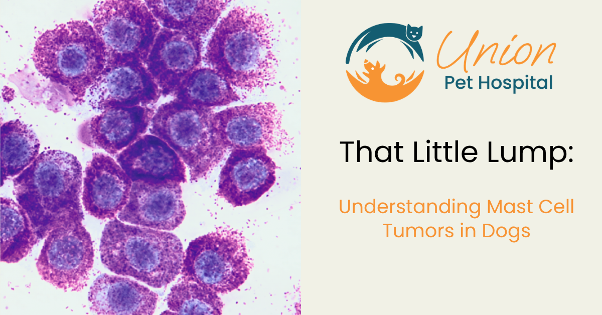
A dog was presented to Union Pet Hospital after their owner noticed a small lump developing on the abdomen. We recommended taking a sample with a needle as the first step in evaluating this new mass, and this showed us mast cells.
That finding changed the conversation from “What is this little lump?” to “What do we need to do next?”
What Is a Mast Cell Tumor in Dogs?
Mast cells are specialized immune cells that are distributed throughout the body’s tissues, particularly near blood vessels and in areas that interface with the environment, such as the skin and gastrointestinal tract. They help regulate inflammation and allergic reactions by releasing substances such as histamine.
A mast cell tumor (MCT) develops when these normally functioning cells begin to grow and divide abnormally, forming a tumor. In dogs, mast cell tumors are the most common type of skin cancer.
One of the challenges with mast cell tumors is that their appearance does not reliably predict their behavior. A tumor may be small and well-defined yet have more aggressive characteristics, while a larger or more irritated mass may ultimately prove to be less aggressive. They also don’t have a consistent appearance. While most are red and raised, they can look and feel like anything.
This is why diagnostic testing is an important part of evaluating any new or changing lump. Cytology from a needle aspirate can often identify mast cells and provide an initial diagnosis of MCT, leading to the recommendation for surgical removal.
Mast cell tumors can also cause changes in the surrounding tissue because of the substances released by the tumor cells. Histamine release, for example, can contribute to swelling, redness, itching, gastrointestinal irritation, and changes in the size or appearance of the mass. This can sometimes make a mast cell tumor appear to change from day to day.
One Lump Doesn’t Always Tell the Whole Story
In this case, the original lump wasn’t the only mass identified. A second mast cell tumor was found while the pet was being prepared for surgery and was removed at the same time.
Both tumors were submitted to the laboratory for histopathology (biopsy), where a pathologist examines each mass individually under a microscope. While an aspirate can help us understand what types of cells are present, biopsy gives us a much more complete picture of the tumor—including its grade and surgical margins.
Two mast cell tumors in the same pet do not necessarily have the same grade or behave in exactly the same way. Each tumor needs to be evaluated on its own.
What Did the Biopsy Show?
The good news in this case was that both tumors were classified as low grade, and the surgical margins were complete. This means the tumors removed during surgery had favorable characteristics and there was no evidence of remaining tumor cells at the examined surgical margins.
Because mast cell tumors can vary from one tumor to another, continued monitoring is still important. The pet’s owners were advised to keep an eye out for any new or changing lumps and to alert their veterinary team if another mass is noticed.
This is an important reminder that even when a tumor has favorable characteristics and has been completely removed, new lumps should still be evaluated rather than assumed to be harmless.
The more we know, the better we can care for your pet.
Why Every Lump Should Be Checked
This case is a good reminder that a lump doesn’t have to look scary to deserve attention.
A small change was noticed and evaluated. Initial testing identified mast cells, which led to surgical removal and submission of the tissue to the laboratory for further evaluation. In this case, both tumors were low grade and completely removed, giving the veterinary team and the pet’s family valuable information about what they were dealing with.
If you find a new lump on your pet, don’t panic—but don’t ignore it either. Your veterinarian can determine whether it should be monitored, sampled, and possibly removed.
Because when it comes to lumps and bumps, knowing is always better than guessing.
What This Case Can Teach Us
🐾 Notice a new lump? Have it checked.
🔬 A needle aspirate can provide valuable information about what cells are present.
🏥 Removal allows the tissue to be submitted for biopsy and more complete evaluation.
📋 For mast cell tumors, grade and surgical margins are important pieces of the puzzle.
👀 Even after a successful removal, watch for new or changing lumps and let your veterinary team know if you find one.
❤️ Early evaluation gives you and your veterinary team the chance to make informed decisions about your pet’s care.
This case is shared for educational purposes. Every pet and every tumor is different, and treatment recommendations should be made with your veterinarian based on your pet’s individual findings.
Medically Reviewed by Dr. Anna Matchinga, DVM
Dr. Matchinga has been with Union Pet Hospital since 2014 and received her Doctor of Veterinary Medicine degree from Auburn University.